Molecular Imaging
Molecular Imaging (MI) is a growing biomedical research discipline that enables the visualization, characterization, and quantification of biologic processes taking place at the cellular molecular level within healthy volunteers and, or patients.
Types of Molecular Imaging
Nuclear Medicine Imaging (PET, SPECT) produces images by detecting signals from distinct parts of the body after a tracer material is injected into the subject. The images are visualized on computer and on media. In nuclear medicine imaging, radiopharmaceuticals are administrated into the body through inhalation, intravenously or orally. External detectors (cameras) capture and form images from the radiation emitted by the radiopharmaceuticals.
There are two main Nuclear Medicine scans (SPECT and PET). Single photon emission computed tomography (SPECT) and positron emission tomography (PET) are nuclear medicine imaging techniques which provide metabolic and functional information unlike more structural CT and MRI machines. PET images are at times combined with CT and MRI to combine anatomical details with metabolic information.
PET affords higher image quality, contrast and tissue resolution compared to SPECT and has higher sensitivity (orders of magnitude higher than SPECT) in addition to being a more quantitative imaging method than SPECT.
Commonly Used Nuclear Medicine Scans for Disease Diagnosis
Bone Density Scan
Enhanced X-ray technology that measures bone loss or density.
Cardiac PET Perfusion
Blood flow (perfusion) to the walls of the heart using a PET scanner. Performed with a cardiac stress test.
Cardiac PET Sarcoid and/or PET Viability
Cardiac PET with different eating instructions. Evaluation of the functional status of the heart for sarcoidosis.
Cardiac SPECT Perfusion
Blood perfusion to the walls of the heart with a cardiac stress test.
PET/CT Scanning
Combined PET/CT scans images of abnormal metabolic activity within the body. The combined scans provide more accurate diagnoses than the two scans separately.
Magnetic Resonance Imaging (MRI)
MRI is a non-invasive imaging technology that produces 3D, detailed corpus images. It is used for disease detection, diagnosis, and treatment monitoring. MRI uses a magnetic field and computer-generated signals to create detailed images of the organs and tissues in the body. MRI machines are circular magnets.
The technology used excites and detects the change in the direction of the rotational axis of protons found in the water that makes up tissue in the body. MRI does not use x-rays or other radiation, so, it is the preferred imaging modality when frequent imaging is required for diagnosis or therapy, especially in the brain.
MRI uses magnets to produce a strong magnetic field that forces protons in the body to match with the magnetic grid. When a current is sent through the patient, protons are stimulated, and are detected by the machine. The MRI sensors can detect the energy released as the protons realign with the magnetic field. Physicians can tell the difference between several types of tissues based on these magnetic properties of the excited tissue.
Diagnostic Ultrasound (US)
Also called sonography or diagnostic echography, is an imaging method that uses sound waves to produce images of structures within your body. The images can provide valuable information for diagnosing and directing treatment for a variety of diseases and conditions. US uses sound waves to produce images of structures within your body. A transducer captures the images. The transducer sends signals into the subject, collects the ones that bounce back and packagers them in a computer, thus creating the image. Ultrasound enables healthcare to visualize the details of soft tissues without making any cuts.
There are three main categories of ultrasound imaging:
- Pregnancy pre-natal ultrasound
- Diagnostic ultrasound
- Ultrasound guided procedures
How Does Molecular Imaging Work?
Most molecular imaging procedures involve an imaging device, a tracer agent, and a detector. A variety of imaging agents are used to visualize cellular activity, including chemical processes like metabolism, oxygenation, and, or blood flow. Imaging agents are typically radiotracers, which is a compound that combines an isotope with a sugar.
Other molecular imaging modalities, such as MRI and molecular ultrasound, use a variety of different agents, typically metallic in origin and polarized. Magnetic resonance (MRI) spectroscopy can measure chemical levels in the body, with or without the use of an imaging agent.
When the imaging agent is introduced into the body, it accumulates in the target organ and, or attaches to specific cells in the body. The imaging camera detects the tracer and creates pictures that show how it is distributed in the body. This distribution pattern helps physicians discern how well organs and tissues are functioning and, or where fluids are flowing and metabolizing in the body.
What is Molecular Imaging Used For?
Molecular imaging is used to diagnose and treat such diseases as Cancer, Alzheimer’s Disease, Parkinson’s Disease, bone disorders, lung disorders, thyroid, and kidney disorders, etc.
-
-
- Shows the extent of the condition of the disease and how it has spread in the body.
- Allows the physician to choose the best and most effective therapy.
- Assesses the patient’s response to certain drugs used for treatment.
- Determines whether a treatment regimen would be effective and, or safe
- Allows for the adaptation of treatment plans when there are changes in cellular activity.
- Assesses disease stages.
-
What is PET Imaging?
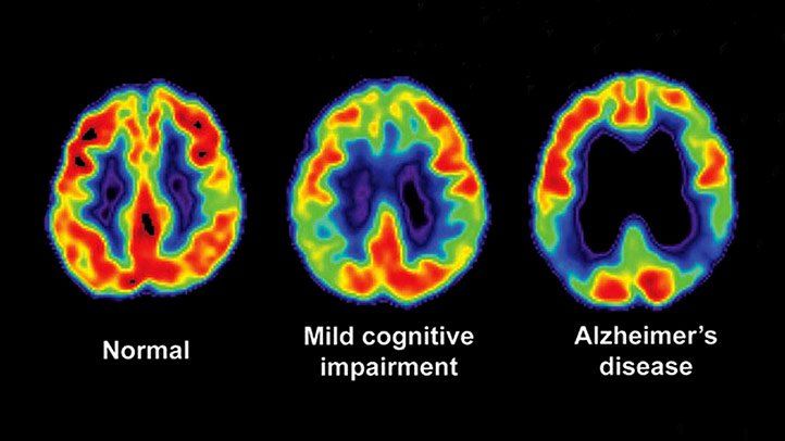
PET involves the use of an imaging device (PET scanner) and a radiotracer that is injected into the patient’s bloodstream. The most used PET radiotracer is 18F-fluorodeoxyglucose (FDG), a compound derived from a simple sugar and a small amount of radioactive fluorine. Once the FDG radiotracer accumulates in the body, it begins to naturally decay and emits positrons that react with the electrons in the human body. This reaction emits energy that is detected by the PET camera.
The PET scanner creates three-dimensional pictures that show how the FDG is distributed around the body. Areas where a large amount of FDG concentrate shows the high level of chemical activity or metabolism to be studied by the researcher or physician. Areas of low metabolic activity appear less intense and are less concerning to the evaluator.
PET-CT is a combination of PET and computed tomography (CT) which produces highly detailed views of the body. The combination of these two imaging techniques allows two different types of scans to be viewed in a single image. CT imaging uses advanced x-ray equipment to produce three-dimensional images. A combined PET-CT study can provide detail on both the structural anatomy and the functional characteristics of organs and tissues.
Neurology and CNS drug development requires PET imaging in combination with CT, MRI, and other imaging technologies to evaluate the metabolic, structural, and chemical safety and efficacy of new drug compounds. The above PET and PET-CT imaging combination technologies will leapfrog CNS therapeutic development in areas of Alzheimer’s, Parkinson’s, Epilepsy, Brain injury and Mental health research.
ADVANTAGES OF MOLECULAR IMAGING IN DRUG DEVELOPMENT
In addition to its role in the diagnosis and treatment of cancer, molecular imaging also serves as a valuable tool in pharmaceutical research and clinical trials. BioPharma (BPSI) is using these additional new imaging techniques including biomarkers to expedite and enhance drug development. Researchers are no longer limited to the use of traditional blood and tissue samples; we can combine traditional pharmacology with molecular imaging.
Molecular imaging is helping researchers gain a better understanding of the basic mechanisms underlying the biology of cells and drug development. Information provided by molecular imaging technologies has the potential to help the drug development and regulatory approval process become faster, more effective, and less costly.
Researchers are also using molecular imaging to identify the most beneficial ways to develop new drugs and to design early phase clinical trials to test their optimum effectiveness.
Researchers have experience in pharmaceutical R&D, collecting pharmacology data, executing imaging programs and pathology studies in the areas of oncology, neuroscience, inflammation, cardiovascular, metabolic, and musculoskeletal, etc.
The physiological and biochemical mechanisms that affect imaging—targets, models, kinetics, and more is well understood by radiology professionals. This vast first-hand experience allows medical professionals to understand drug behavior and translate knowledge to conduct effective studies and gather useful data.
Imaging, as an assay that enables visualization of subcellular processes and diagnostic of clinical imaging biomarkers, including everything in between, has become a new weapon in the drug development arsenal. With all the imaging techniques that exist, researchers use medical methods to ensure data is quantifiable, accurate and valid.
This is all to ensure that the results can be used to drive meaningful decisions. Center of Excellencies like Washington University in St Louis, Johns Hopkins, Ktatholieke Universiteit Leuven University are leading the way in Molecular Imaging solutions to aid in Phase 1 Research with Phase 1 Clinics like BioPharma Services in Toronto.
Ready To Get Started?
Complete the Form below to schedule a Discovery Call with a member of our medical team to learn how BioPharma Services can be your trusted Nuclear Imaging partner!
Schedule a Discovery Call
You can unsubscribe at any time. For more details, please read our Privacy Policy.

